סריקה בתהליך...
הניתוח עשוי לארוך 30–90 שניות
הידעת?
הסרטון אינו מכיל תוכן רפואי
חושבים שחלה טעות?
אם לדעתכם הסרטון אכן עוסק בטענות רפואיות או בריאותיות, הוסיפו הסבר קצר ושלחו בקשה לבדיקה מחדש.
הבקשה התקבלה! נבדוק את הסרטון ונחזור אליכם.
אירעה שגיאה בשליחת הבקשה. נסו שוב.
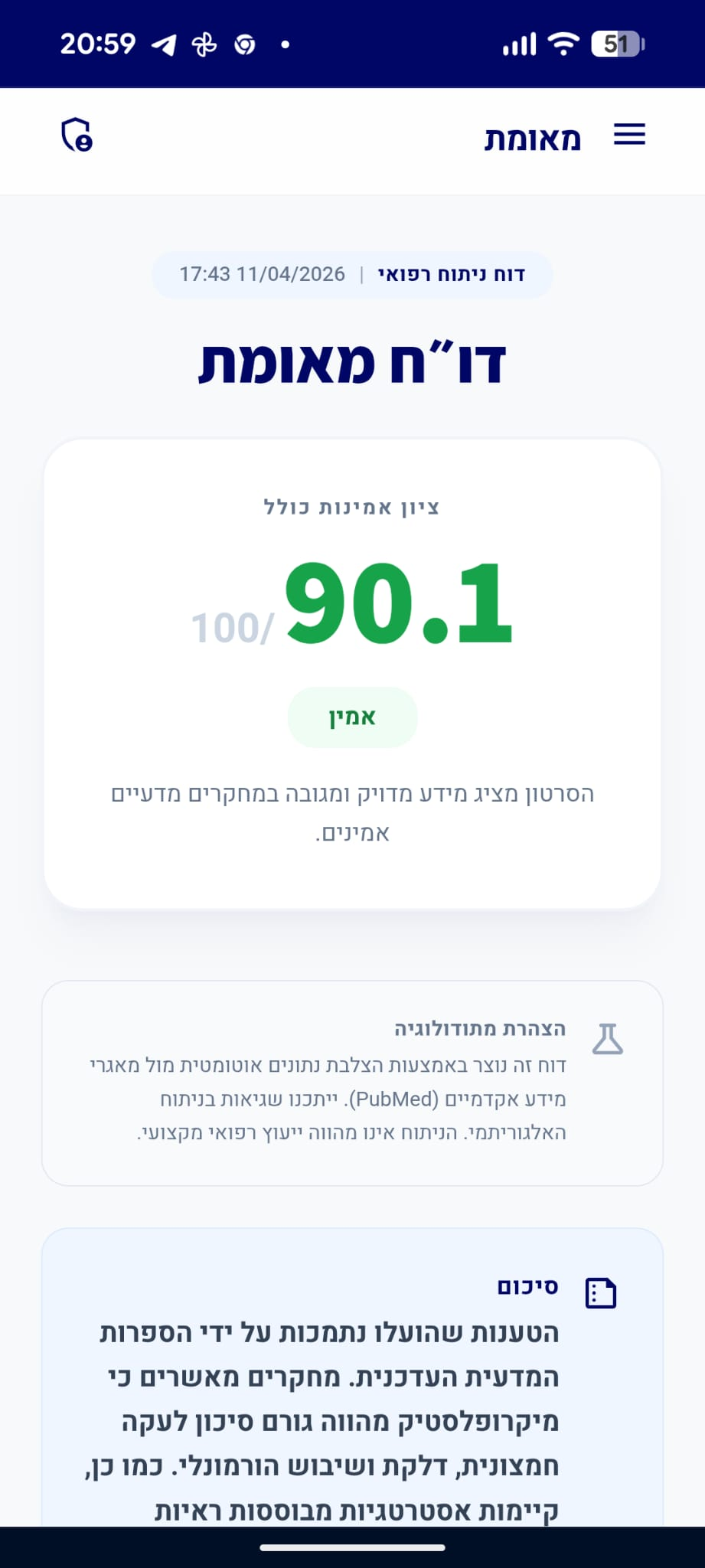
דו״ח מאומת
הסרטון מציג מידע מדויק ומגובה במחקרים מדעיים אמינים.
סיכום
כל הטענות שהועלו נתמכות באופן חד-משמעי על ידי הספרות המדעית העדכנית ב-PubMed. קיימת הסכמה רחבה בקהילה הרפואית לגבי חשיבות צריכת הנוזלים, תפקידו המגן של הציטראט, והיותן של אבני סידן אוקסלאט הסוג הנפוץ ביותר.
analytics ניתוח טענות מבוסס ראיות
"פירות הדר, כגון תפוזים ולימונים, עשויים לסייע במניעת אבנים בכליות."
מסקנת הבדיקה:
מחקרים אפידמיולוגיים וסקירות שיטתיות מצביעים על כך שצריכת פירות הדר ומיציהם, המכילים ציטראט, קשורה לירידה בסיכון להיווצרות אבנים בכליות. הציטראט פועל כמעכב טבעי של התגבשות מלחי סידן בשתן. (🟩)
chevron_right מקורות מדעיים: (2)
-
link
Role of Citrus Fruit Juices in Prevention of Kidney Stone Disease (KSD): A Narrative Review.
To explore the relationship between citrus fruit juices (oranges, grapefruits, and lemonades) and kidney stone disease (KSD). A systematic review was performed using the Medline, EMBASE, and Scopus databases, in concordance with the PRISMA checklist for all English, French, and Spanish language studies regarding the consumption of citrus fruit juices and the relationship to urinary stone disease. The main outcome of interest was the association of citrus fruit juices with KSD. Thirteen articles met the criteria for inclusion in the final review. Three large epidemiological studies found that grapefruit juice was a risk factor for stone formation, while orange juice did not increase the risk for KSD. Ten small prospective clinical studies found that orange, grapefruit, and lemon juices all increased urinary citrate levels. Only orange and grapefruit juices had an alkalinizing effect and while lemon juice has a protective effect by raising urinary citrate levels, it lacked a significant alkalinizing effect on urine pH. Orange juice and grapefruit juices significantly increased urinary oxalate levels, while orange juice also had a high carbohydrate content. While orange juice seems to play a protective role against stone formation, grapefruit was found to raise the risk of KSD in epidemiological studies but had a protective role in smaller clinical studies. Lemon juice had a smaller protective role than orange juice. Larger amounts of, as well as more accurate, data is needed before recommendations can be made and a high carbohydrate content in these juices needs to be taken into consideration.…
PMID: 34836376
-
link
Associations between comprehensive dietary composition and kidney stone risk: insights from a nationally representative survey.
<h4>Background</h4>This study aimed to identify key dietary components exhibiting significant associations with risk of kidney stone (KS).<h4>Methods</h4>This is a cross-sectional analysis that included data from the National Health and Nutrition Examination Survey (2007-2020) based on 26,372 qualified individuals, who provided self-reported information regarding KS and dietary composition over two days. The relationship between the risk of KS and dietary composition were evaluated using weighted multivariate logistic regression models and restricted cubic spline (RCS) models.<h4>Results</h4>Through weighted multivariate logistic regression model, daily consumption of citrus, melons, and berries (OR = 0.81, 95% CI = 0.70-0.93), tomatoes (OR = 0.78, 95% CI = 0.61-0.99), milk (OR = 0.84, 95% CI = 0.78-0.91), total dairy (OR = 0.89, 95% CI = 0.84-0.94), and alcoholic drinks (OR = 0.88, 95% CI = 0.84-0.92) were associated with a lower likelihood of KS development, while daily consumption of added sugars correlated with an elevated probability of KS occurrence (OR = 1.01, 95% CI = 1.00-1.01). Restricted cubic spline analysis found that total fruits, total vegetables, total protein foods, total grains, total dairy, oils, solid fats and added sugars and the risk of KS were in a curvilinear relationship adjusted for age, sex, race, marital status, BMI (body mass index), physical activity in recreational time, smoking status, hypertension, diabetes mellitus (DM). (p overall and p non-linear <0.05).<h4>Conclusion</h4>This cross-sectional study elucidates intricate curvilinear associations between dietary components and the risk of KS. The consumption of citrus, melons, berries, tomatoes and milk was associated with a significantly lower risk of KS. Our findings highlight the need for prospective studies to confirm these potential protective relationships.…
PMID: 41245408
"אבני סידן אוקסלאט הן הסוג הנפוץ ביותר של אבנים בכליות."
מסקנת הבדיקה:
הספרות הרפואית המקובלת מאשרת כי אבני סידן אוקסלאט הן הסוג הנפוץ ביותר של אבנים בדרכי השתן, ומהוות מעל 80% מכלל המקרים. (🟩)
chevron_right מקורות מדעיים: (2)
-
link
Urolithiasis: History, epidemiology, aetiologic factors and management.
Urolithiasis is defined as a disease diagnosed by the presence of one or more stones in the urinary tract. It is one of the oldest and most widespread diseases known to man, their discovery and characterisation chronology began with the civilisation's history. This pathology has a multifactorial aetiology, very frequent worldwide with geographic and racial variation, their prevalence is increasing in lockstep with socioeconomic development. In fact, this disorder affects between 2 and 20% of the population, with an approximate recurrence rate of 30% to 50% in 5 years. Furthermore, calciumtype stones, which are composed of calcium oxalate (CaOx) alone or a mixture of CaOx and calcium phosphate are the most common, accounting for more than 80% of cases. The medical management of urolithiasis is done by medical treatments and/or by surgical intervention for the stones extraction by the techniques such as extracorporeal shock wave lithotripsy (ESWL), ureteroscopy (URS), percutaneous nephrolithotomy (PCNL) and open surgery. However, various therapies, including thiazide diuretics and alkaline citrate, are used in an attempt to prevent stones recurrence induced by hypercalciuria and hyperoxaluria, but the scientific evidence for their effectiveness is less convincing. On the other hand, endoscopic and ESWL methods have revolutionised the treatment of urinary lithiasis, but these costly methods, can cause acute kidney injury and decreased renal function, in addition, do not prevent the probability of new stone formation. The deepening of our knowledge on all points relating to this disease is a priority for specialists in order to find adequate solutions for this disease. This review provides an overview of urolithiasis, its history, epidemiology, clinical manifestation, diagnosis and treatment methods.…
PMID: 38155376
-
link
Kidney stones.
Kidney stones are mineral deposits in the renal calyces and pelvis that are found free or attached to the renal papillae. They contain crystalline and organic components and are formed when the urine becomes supersaturated with respect to a mineral. Calcium oxalate is the main constituent of most stones, many of which form on a foundation of calcium phosphate called Randall's plaques, which are present on the renal papillary surface. Stone formation is highly prevalent, with rates of up to 14.8% and increasing, and a recurrence rate of up to 50% within the first 5 years of the initial stone episode. Obesity, diabetes, hypertension and metabolic syndrome are considered risk factors for stone formation, which, in turn, can lead to hypertension, chronic kidney disease and end-stage renal disease. Management of symptomatic kidney stones has evolved from open surgical lithotomy to minimally invasive endourological treatments leading to a reduction in patient morbidity, improved stone-free rates and better quality of life. Prevention of recurrence requires behavioural and nutritional interventions, as well as pharmacological treatments that are specific for the type of stone. There is a great need for recurrence prevention that requires a better understanding of the mechanisms involved in stone formation to facilitate the development of more-effective drugs.…
PMID: 27188687
"ציטראט נקשר לסידן בשתן ובכך מונע ממנו להתחבר לאוקסלאט וליצור גבישים."
מסקנת הבדיקה:
ציטראט בשתן נקשר ליוני סידן ויוצר קומפלקס מסיס, ובכך מפחית את זמינות הסידן להיקשרות עם אוקסלאט. תהליך זה מונע את היווצרותם של גבישי סידן אוקסלאט. (🟩)
chevron_right מקורות מדעיים: (2)
-
link
Calcium Phosphate Nephrolithiasis: A Comprehensive Review.
In this article, we review the recent epidemiology, unique pathophysiology, and challenges in medical management of calcium phosphate (CaP) kidney stones. CaP stones represent the second most encountered stone type. Compared with the more common calcium oxalate stones, CaP stones are more likely to occur in women, have increased in prevalence in recent decades, and recur at a higher rate. Stone formers presenting with hydroxyapatite and brushite stones, the 2 most common subtypes of CaP stones, exhibit distinct histopathologic findings. Urinary risk factors contributing to CaP stone formation include high urine pH, hypercalciuria, and hypocitraturia. These changes in the urinary environment occur from a variety of inherited or acquired conditions. The current approach to medical management of CaP stones is primarily extrapolated from studies performed in calcium oxalate stone formers, and the role of alkali therapy in CaP stone formers is controversial. Therefore, there is a critical need for treatments tailored to address the high recurrence rate, distinct pathophysiology, and risk factors of CaP nephrolithiasis.…
PMID: 41940378
-
link
Glycine suppresses kidney calcium oxalate crystal depositions via regulating urinary excretions of oxalate and citrate.
An abnormal urine composition is a key reason for kidney stone formation, but little is known about the roles of small metabolites in the urine during kidney stone formation. Here, we found urine glycine in patients with kidney calcium oxalate (CaOx) stone was significantly lower than that in healthy people via…
PMID: 33772775
"טיפול ב-Potassium Citrate הוא טיפול מניעתי מקובל לחלק מהמטופלים הסובלים מאבנים חוזרות בכליות."
מסקנת הבדיקה:
טיפול תרופתי ב-Potassium Citrate הוא אסטרטגיה קלינית מקובלת למניעת הישנות אבנים, במיוחד אצל מטופלים הסובלים מהיפוקיטרטוריה (רמות ציטראט נמוכות בשתן). (🟩)
chevron_right מקורות מדעיים: (2)
-
link
Calcium Phosphate Nephrolithiasis: A Comprehensive Review.
In this article, we review the recent epidemiology, unique pathophysiology, and challenges in medical management of calcium phosphate (CaP) kidney stones. CaP stones represent the second most encountered stone type. Compared with the more common calcium oxalate stones, CaP stones are more likely to occur in women, have increased in prevalence in recent decades, and recur at a higher rate. Stone formers presenting with hydroxyapatite and brushite stones, the 2 most common subtypes of CaP stones, exhibit distinct histopathologic findings. Urinary risk factors contributing to CaP stone formation include high urine pH, hypercalciuria, and hypocitraturia. These changes in the urinary environment occur from a variety of inherited or acquired conditions. The current approach to medical management of CaP stones is primarily extrapolated from studies performed in calcium oxalate stone formers, and the role of alkali therapy in CaP stone formers is controversial. Therefore, there is a critical need for treatments tailored to address the high recurrence rate, distinct pathophysiology, and risk factors of CaP nephrolithiasis.…
PMID: 41940378
-
link
Management of urinary stones by experts in stone disease (ESD 2025).
The formation of kidney stones is a complex biologic process involving interactions among genetic, anatomic, dietary, and environmental factors. Traditional lithogenic models were based on urine supersaturation in relation to the activity of crystallization promoters and inhibitors. However, modern research has added new principles such as the "renal epithelial cell response" and the role of inflammation and oxidative stress leading to the development of a "multi-hit hypothesis". A strong correlation between urinary stones and kidney damage has been well demonstrated by both cohort and case-control studies. The main contributors to chronic kidney damage associated with urinary stones include crystal deposition within the renal parenchyma, associated comorbidities, repeated obstructive and infectious episodes, as well as the potential adverse effects of stone removal procedures. Most hereditary stones may cause high urinary saturation levels promoting obstruction of the Bellini ducts and consequent glomerulosclerosis and interstitial fibrosis in the cortex. These include hereditary hypercalciurias, primary hyperoxalurias, cystinuria, adenine phosphoribosyltransferase (APRT) deficiency (associated with 2,8-dihydroxyadenine lithiasis) and xanthinuria. Complete distal renal tubular acidosis occurs in childhood and presents deafness, rickets, and a short life expectancy. The incomplete form usually manifests in adulthood, primarily with recurrent urinary lithiasis, and less frequently with nephrocalcinosis. In all stone formers stone analysis and a basic metabolic evaluation, including blood biochemistry, urine sediment examination, urinary pH and culture are mandatory, in contrast high-risk stone formers require a more specific metabolic evaluation, including a 24-hour urine sample to measure calcium, phosphate, citrate, oxalate, uric acid, magnesium, sodium and proteinuria. The morpho compositional analysis of kidney stones offers essential insights beyond merely identifying their predominant chemical component. This approach reveals key aspets of the stone formation, such as nucleation sites, crystal growth patterns, and the presence of specific lithogenic processes. The ideal analytical protocol combines stereoscopic microscopy (StM), scanning electron microscopy with energy-dispersive X-ray spectroscopy (SEM-EDS), and, when necessary, Fourier-transform infrared spectroscopy (FTIR). Recurrence prevention and managing residual fragments require complementary strategies such as lifestyle modifications, dietary interventions, and pharmacological therapies. Among pharmacological options, alkaline citrate salts, particularly potassium citrate, are widely used due to their ability to modify urinary chemistry and inhibit stone formation. Recently, novel molecules have been introduced into the management of renal stone disease. Phytate a naturally occurring polyphosphorylated carbohydrate, exibits a potent inhibitory effect on calcium salt's nucleation, growth, and aggregation. Theobromine, another natural compound, has been shown to effectively inhibit uric acid crystallization. The co-administration of urinary alkalinizing agents, such as potassium citrate, alongside theobromine has been proposed as a therapeutic strategy to optimize uric acid solubility and to reduce the risk of excessive alkalinization and subsequent sodium urate precipitation. Struvite stones are caused by urinary tract infection with urease- producing microorganisms. Their treatment requires specific measures including complete surgical stone removal, short or long-term antibiotic treatment, to maintain urinary acidification to a pH below 6.2, and a urine volume of at least 2 litres/24 hours. L-methionine has been shown to effectively lower urine pH and the relative supersaturation of struvite. An essential aspect of medical management of urinary stone disease is treatment adherence, which depends on perceived benefit, treatment duration, and side effect profile. The side effects of citrate treatment are mild gastrointestinal disorders whereas thiazide diuretics tend to cause hypokalemia-related symptoms and less frequent metabolic and dermatologic side effects. Urease inhibitors for struvite stones and drugs used to enhance cystine solubility are more frequently associated with side effects. The use of smartphone applications can support patients by promoting adequate hydration, adherence to dietary recommendations, and compliance with prophylactic medication. Endoscopic techniques currently play a prevalent role in the removal of renal stones, while extracorporeal shock wave lithotripsy is today marginally used for specific indications. Different technical modalities can be used for percutaneous nephrolithotomy (PCNL), each with its own advantages and disadvntages (standard vs. mini, prone vs. supine, fluoroscopic vs ultrasound-guided). Flexible ureteroscopy or retrograde intrarenal renal surgery (RIRS) has extended its indications due to technological advancements in endoscopes and their accessories. The availability of new laser technologies (thulium fiber laser and pulse-modulated Ho:YAG laser) has enhanced stone fragmentation and dusting capabilities. However, their use exposes the renal parenchyma to high temperatures and pressures which could potentially contribute to renal damage. Factors influencing heat release include laser type and settings, exposure time, stone location, fiber-to-stone distance, irrigation volume and fluid circulation. Reduction of heat release can be achieved by limiting the laser settings to reasonable values or by improving fluid circulation with use of ureteral access sheaths, especially those navigable and equipped with suction. High intrarenal pressure is also closely associated with renal damage. Sustained high pressure or even pressure spikes may increase this risk, highlighting the importance of real-time pressure monitoring through sensors integrated on guidewires, scopes, access sheath and use of innovative platforms regulating irrigation/suction systems. Direct In-Scope Suction (DISS) system was developed to control intrarenal pressure and facilitate the removal of residual fragments. Flexible and Navigable Suction Ureteral Access Sheath (FANS-UAS) is a flexi-bendable UAS equipped with suction capabilities combining mechanical flexibility with continuous irrigation management and stone clearance mechanisms. Ultra-thin scopes (7.5 F) make it easy to perform RIRS without the need for pre-placed double-J stents or with a 9 F sheath achieving more space for stone fragments expulsion or infusion. All these technological advancements have enhanced the efficacy of fURS or RIRS which can be an alternative treatment (salvage fURS) when standard stone management techniques, such as percutaneous nephrolithotomy (PCNL), are contraindicated or fail. Salvage fURS has shown favorable outcomes in complex or high-risk cases, including patients with coagulopathies, morbid obesity, renal anatomical abnormalities (e.g., horseshoe or pelvic kidneys), urinary diversion, calyceal diverticula, and altered urinary tracts. In such scenarios it demonstrated favorable outcomes with stone-free rates ranging from 55.6% to 64% for stones > 2 cm. Although non-invasive, extracorporeal and endoscopic treatments for renal and ureteral stones carry a risk of complications that can be classified according to the Clavien-Dindo system. The complication rate after SWL was estimated at 18.43% for Clavien grade I-II complications (pain, hematuria) and 2.48% for Clavien III-IV complications (hematoma, sepsis). The most frequent complication after RIRS is fever or urinary tract infection observed in 0.2-15% (with 0.1-4.3% of cases of urinary sepsis). Complications after PCNL are more frequent and may include moderate events (hemorrhage requiring transfusion 2-7%, urosepsis 1-2%, bowel injury < 1%) as well as severe events (arteriovenous fistula 0.5-1%, thoracic complications < 1% , loss of access tract 1-3%, death < 0.5%). The risk of bleeding complications is significantly increased in patients on antithrombotic therapy. A personalized, interdisciplinary approach enables optimal decision-making in balancing antithrombotic therapy with surgical safety during urological stone interventions Finally, it must be considered that endourological procedures can be harmful to the surgeons themselves and their team due to exposure to ionizing radiation. For this reason, procedures must be carried out in strict accordance with safety guidelines and regulations to minimize radiation exposure. Safety is vital in any surgical intervention, with efficacy being the next most critical consideration. However, cost-effectiveness should be also considered. Endourology involves high costs largely due to the use of sophisticated equipment that requires frequent renewal due to the continuous rapid technological evolution. Using disposable devices brings numerous benefits but also leads to a further increase in costs. Finally, in the cost-benefit assessment, the rate of reintervention associated with some types of procedures must be considered.…
PMID: 40583613
"שתייה מספקת של מים היא אחד הגורמים החשובים ביותר במניעת אבנים בכליות."
מסקנת הבדיקה:
צריכת נוזלים מספקת נחשבת לגורם המניעתי החשוב ביותר באבנים בכליות, שכן היא מדללת את ריכוז המלחים בשתן ומונעת את תהליך ההתגבשות. (🟩)
chevron_right מקורות מדעיים: (3)
-
link
Dietetic and lifestyle recommendations for stone formers.
Nutrition is tightly associated with the risk of stone events. A part from genetic predisposition, a correct and balanced diet might prevent incident kidney stones. Several studies analyzed each dietary component and different diets to better understand their impact on stone recurrence. Fluids: High fluids intake is the most important factor for preventing kidney stones disease and for every 200 mL of water, the risk of stones is reduced by 13%. Soft drinks seems to be associated to a greater risk of stone events, whereas caffeine and citrus fruits juice are not. Calcium: Normally calcium intake with diet does not exceed 1.2 g/day. A balanced consumption of dairy products is capable of reducing oxalate intestinal absorption and urinary excretion compared to low calcium diet, being protective for stone disease. Oxalate: The exact amount of oxalate contained in different foods is difficult to estimate for its variability, even in the same aliment. In addition, the amount of oxalate consumed was shown to be only a minor risk factor for stone disease, whereas its intestinal absorption is strongly influenced by external factors, such as calcium intake. Dietary oxalate restriction is advisable only in patients with known elevated consumption. Sodium: High sodium intake is both associated with hypertension, heart disease and stone risk. Increased sodium consumption is directly associated to hypercalciuria in both calcium stone formers and healthy subjects. Although dietary sodium restriction to recommended values is always desirable in stone formers, it is difficult to achieve for its broad use in food preparation. Proteins: Animal proteins are associated to increased risk for stone formation, whereas vegetable and dairy proteins are not. Increased meat intake was associated to acidic urine pH, negative calcium balance and reduced anti-lithogenic urinary solutes excretion.Fruits and vegetables: Alkalizing foods are one of the most important factors for stone protection. Their consumption increases anti-lithogenic solutes as citrate, potassium and magnesium. A diet rich in fruits and vegetables is strongly recommended for stone formers. Uric acid: Elevated meat consumption is either associated to increased purine metabolism and acid load, favoring uric acid nephrolithiasis by reducing urine pH and increasing urinary excretion of uric acid, especially in patients affected by metabolic syndrome and diabetes.In conclusion, the most effective diet for stone protection is rich in fruits and vegetables, low in animal proteins and salt, with balanced dairy product consumption and obviously, with elevated fluid intake. These characteristics make vegetarian and Mediterranean diets protective and useful for stone formers, whereas western diet is at risk for stone formation. La nutrición está fuertemente asociada al riesgo de episodios litiásicos. Aparte de la predisposición genética, una dieta correcta y balanceada podría prevenir la incidencia de litiasis renal. Varios estudios han analizado cada componente de la dieta y diferentes dietas para entender mejor su impacto sobre la recurrencia litiásica. Líquidos: Una alta ingesta de fluidos es el factor más importante para la prevención de la enfermedad litiásicay por cada 200 mL de agua, el riesgo de litiasis se reduce un 13%. Los refrescos parecen estar asociados a un mayor riesgo de eventos litiásicos, mientras que la cafeina y los zumos de cítricos no lo están. Calcio: Normalmente la ingesta diaria de calcio con la dieta no excede los 1,2 g. Un consumo balanceado de productos lácteos es capaz de reducir la absorción intestinal y la excreción urinaria de oxalato si lo comparamos con una dieta pobre en calcio, siendo protector para la enfermedad litiásica. Oxalato: La cantidad exacta de oxalato en las diferentes comidas es difícil de estimar debido a su variabilidad, incluso en el mismo alimento. Además, se demostró que la cantidad de oxalato ingerido era solo un factor de riesgo menor para la enfermedad litiásica, mientras que su absorción intestinal está fuertemente influenciada por factores externos como la ingesta de calcio. La restricción de oxalato en la dieta se aconseja solamente en pacientes con aumento probado en su consumo. Sodio: Una ingesta elevada de sodio se asocia tanto con la hipertensión como con la enfermedad cardiaca y el riesgo de litiasis. El consumo elevado de sodio está directamente asociado con la hipercalciuria, tanto en litiásicos cálcicos como en sujetos sanos. Aunque la restricción dietética de sodio a los valores recomendadoses deseable en los pacientes litiásicos, es difícil de conseguir debido al alto uso de sodio en la preparación de las comidas.Proteinas: Las proteinas animales están asociadas a un riesgo aumentado para la formación de litiasis, mientras que las vegetales y las de productos lácteos no. Un aumento de consumo de carne se ha asociado a un pH urinario ácido, balance de calcio negativo y reducciónde la excrección urinaria de solutos anti-litogénicos.Frutas y vegetales: Las comidas alcalinizantes son uno de los factores más importantes para la protección ante la litiasis. Su consumo aumenta los solutos anti-litogénicos como el citrato, potasio y magnesio. Una dieta rica en frutas y vegetales es muy recomendable para los pacientes litiásicos. Acido úrico: Un consumo elevado de carne está asociado tanto a un aumento de metabolismo de las purinas como a la carga ácida, favoreciendo la nefrolitiasis úrica al reducir el pH urinario y aumentar la excreción urinaria de ácido úrico, especialmente en pacientes afectados por el síndrome metabólico y diabetes.En conclusión, la dieta más efectiva para proteger contrala litiasis es la rica en frutas y vegetales, pobre en proteinas animales y sal, con un consumo balanceado de productos lácteos y, obviamente, con una alta ingesta de líquidos. Estas características hacen que las dietas vegetarianas y la mediterránea sean protectoras y útiles para los pacientes litiásicos, mientras que la dieta occidental sea una dieta de riesgo para la formación de litiasis.…
PMID: 33459627
-
link
Nutrition and Kidney Stone Disease.
The prevalence of kidney stone disease is increasing worldwide. The recurrence rate of urinary stones is estimated to be up to 50%. Nephrolithiasis is associated with increased risk of chronic and end stage kidney disease. Diet composition is considered to play a crucial role in urinary stone formation. There is strong evidence that an inadequate fluid intake is the major dietary risk factor for urolithiasis. While the benefit of high fluid intake has been confirmed, the effect of different beverages, such as tap water, mineral water, fruit juices, soft drinks, tea and coffee, are debated. Other nutritional factors, including dietary protein, carbohydrates, oxalate, calcium and sodium chloride can also modulate the urinary risk profile and contribute to the risk of kidney stone formation. The assessment of nutritional risk factors is an essential component in the specific dietary therapy of kidney stone patients. An appropriate dietary intervention can contribute to the effective prevention of recurrent stones and reduce the burden of invasive surgical procedures for the treatment of urinary stone disease. This narrative review has intended to provide a comprehensive and updated overview on the role of nutrition and diet in kidney stone disease.…
PMID: 34204863
-
link
The role of fluid intake in the prevention of kidney stone disease: A systematic review over the last two decades.
The incidence of kidney stone disease (KSD) is rising worldwide; hence, more focus must be directed toward its etiology and risk factors. Increasing fluid intake is recommended as the most ideal prevention; yet, there is inconsistent evidence surrounding optimum volumes and types of fluid that affect stone formation. This review aimed to analyze the published literature on fluid intake and types of fluid consumed and their impact on KSD prevention. Papers were acquired from databases: MEDLINE, EMBASE, PubMed, CINAHL, and Cochrane Library. Included English language studies that involved adults consuming beverages along with a standardized diet in relation to KSD. Those failing to control dietary factors were excluded. After an initial search of 1099 papers, 9 (541 participants) were included in the final review. Six varieties of water and ten different types of juices were investigated. Higher fluid intake was associated with increased urine output and reduced stone formation. Water with high calcium content seemingly increased the rate of calcium oxalate (CaOx) stone formation. The relative supersaturation of CaOx in urine was decreased with grapefruit, apple, orange juices, and sodas, whereas cranberry juice increased it. Plum juice and the energy drink Gatorade had no effect on stone formation. Fluids low in calcium seem to reduce the risk of KSD. Certain varieties of fluid, such as grapefruit, apple, and orange juices reduce urine CaOx saturation, with a subsequent reduction in stone formation. Findings from this review could contribute to primary prevention for those at risk of KSD.…
PMID: 32525478

Lior Ben Moshe | רפואה בשפה פשוטה
דירוג זה מבוסס על 2 דוחות אימות קודמים.

האם הדוח הזה היה מועיל לך?
מה היה פחות טוב? (רשות)
תודה על הפידבק!
עירעור על דוח זה
ספקו ראיות חדשות או הצביעו על אי דיוקים
נעדכן אותך על תוצאות הבדיקה
הוסיפו קישורים למחקרים או מקורות רפואיים מוכרים
העירעור נשלח בהצלחה!
המנוע המדעי שלנו יבדוק את הראיות שהגשתם. נעדכן אתכם באימייל עם התוצאות.
ניתוח מבוסס בינה מלאכותית
דוח זה נוצר באופן אוטומטי על ידי מערכת בינה מלאכותית ועשוי להכיל שגיאות, אי-דיוקים או מידע חלקי. הניתוח אינו מהווה ייעוץ רפואי, אבחנה או המלצה לטיפול, והוא אינו תחליף לדעתו של איש מקצוע רפואי מוסמך. יש להתייעץ עם רופא או מומחה מוסמך לפני קבלת כל החלטה רפואית. המידע מוצג לצרכי מידע כללי בלבד.
מידע זה מופק על ידי בינה מלאכותית ואינו מהווה תחליף לייעוץ רפואי מקצועי.